Monday, September 30, 2013

When should I seek Medical Treatment for foot Pain?


When should I seek medical treatment for foot pain?


When the pain begins to interfere with your activities of daily living or if you cannot perform your desired activities without pain, you should consider seeking medical attention. Indicators that you should seek medical care are if the area looks deformed, you have loss of function, change of sensation, a large amount of swelling with pain, prolonged change of skin or toenail color, the affected area becomes warmer than the surrounding areas, becomes exquisitely tender to the touch, or is causing you to move differently.

How is foot pain diagnosed?


Proper evaluation and diagnosis of foot pain is essential in planning a treatment. A good general guideline is to compare the injured side to the uninjured side. Injury may present itself as distinguishable lump or gap felt at that location or a "crunchy" feeling on that spot caused by inflammation. The types, causes, and severity (sharp pain versus a dull ache) also are good indicators of the seriousness of the injury.

  • Four grades of pain
    • During activity
    • Before and after, and not affecting performance
    • Before, during, and after athletic activity, affecting performance
    • Pain so severe that performance is impossible
The doctor will ask you several questions to determine how the problem began. It can be helpful to tell the physician about how and when it started, how it affects you, when it bothers you, what you may or may not have done to make the pain better or worse. If necessary, a thorough physical exam may be conducted to evaluate for any other injuries.
  • Feet will be physically and visually examined at rest, with weight- and non-weight-bearing movement by the medical professional.
  • The foot and arch will be touched and manipulated and inspected to identify obvious deformities, tender spots, or any differences in the bones of the foot and arch.
  • Dr. Raymond A. DiPretoro, Jr. will examine how the muscles of your foot function. These tests may involve holding or moving your foot and ankle against resistance, you may also asked to stand, walk, or even run.
  • The nerves in the foot will be tested to make sure no injury has occurred there.
  • An X-ray, MRI, or bone scan of the foot and arch may be taken to determine if there are abnormalities of the bone and/or soft tissues.

Sunday, September 29, 2013

How can foot Pain be Prevented?


How can foot pain be prevented?


Physicians with sports medicine, physical medicine, or orthopedic backgrounds may also help you choose an appropriate activity. After choosing the sport or activity that you wish to participate in, proper preparation will help minimize the initial aches and pains of that activity. Proper technique in any activity will help you to properly and safely perform your chosen activity and avoid injury. Good coaching can help you develop good biomechanics that can prevent foot pain.To prevent injuries and pain, the following issues should be addressed before starting an exercise routine. Are you in good health? A general physical exam by a physician will help to evaluate your cardiovascular function, the possibility of disease or any other general medical problems that you may have. Before beginning activities, diseases such as gout, diabetes, certain types of arthritis, and neuropathies should be treated.
Shoes and socks appropriate to your activity will also be a deterrent to foot pain. Properly fitting shoes and proper foot hygiene can prevent blisters, ingrown toenails, corns, calluses, bunions, stress fractures, metatarsalgia,Morton's neuroma, mallet toes, and plantar fasciitis. Poorly fitting footwear can make poor biomechanics worse, and properly fitting footwear can help to minimize the effect of bad biomechanics.
A plan for a gradual return to play should be started once the pain is reduced and muscle strength and flexibility are restored. Returning to participation and prevention of foot pain are governed by the same factors as preparing for participation. Foot pain can be caused by doing too much of a particular activity too fast. Ignoring pain can also lead to further problems with the foot. Different types of foot pain can be seen at different times of the season. Typically, blisters, shin splints, and arch injuries are seen at the beginning of the season. Again, to avoid blistering in the future, a generous application of petroleum jelly to the affected area can be helpful.
Stress-related problems are related to the workloads. If the body is not prepared for an increase of workload that is typical early in the season and with "weekend warriors," acute shin splints and tendonitis are very common, in addition to increased muscle soreness.
After one has foot pain, an optimal workout program begins with a physical exam by a physician, followed by a gradual, consistent workout plan. A good example of this type of program is a running program that starts with a good warm-up, such as walking five to 10 minutes, then alternating sets of jogging and walking. An example of such a program would be 20 sets of jogging for two minutes, then walking one minute, with jogging time increased until you can run continuously for 40 minutes. Good surfaces and proper equipment used in your workout will lower the risk of foot pain.
Components of a good exercise program should include core strengthening, muscle strengthening, and flexibility specific to the goals of the workout program or the sport.
If pain is encountered when working out, try decreasing the intensity of the workout. If the pain persists, then you should immediately stop and seek medical advice from your Podiatrist at Advanced Foot & Ankle Center to discover the source of the pain. Pushing through pain often results in injury.

Thursday, September 26, 2013

What other symptoms and signs accompany foot pain?


What other symptoms and signs may accompany foot pain?


Pain and point tenderness are the immediate indicators that something is wrong in a specific area. The onset of pain, whether suddenly or over time, is an important indicator of the cause of the problem. The following questions are also important.

  • Is there pain with movement of the affected area?
  • Is it affected by weight-bearing?
  • Does it change your walking motion?
Bones of the foot are joined together by ligaments. A sprain occurs when the ligaments that hold the bones together are overstretched and the fibers tear. Point tenderness and looseness of a joint can be indicators of a sprain.
Injury to the bones of the foot can be caused by a single blow or twist to the arch or also by repetitive trauma that can result in a stress fracture. Fractures are indicated by a focal point of pain that may be exquisitely tender on the bone. There may be a distinguishable lump or gap at the site of the fracture. A rotated toe or forefoot may also be a sign of a fracture or dislocation.
Muscle and tendons move the body tissues around the joints. A strain occurs when a muscle or group of muscles are stressed to the point where there is tearing of the muscle fibers. The muscles and tendons of the foot may be strained by overstretching, overuse, overloading, bruising, or even being lacerated. Weakness in contraction of a joint, difficulty in stabilizing body parts, and pain working against resistance are signs of muscle problems. Swelling, tenderness, loss of function, and discoloration over and around the injury of can be symptoms and signs of a strain.
Bruises (contusions) are most commonly the result of a direct impact injury to the body. A bruise can occur to the foot by a variety of causes, such as having your foot stepped on or by stepping on a rock. Blows to the foot that result in pain, discoloration, swelling, and changes in how you walk may indicate more serious damage such as fractured bone.
Pain and tenderness associated with planter fascia strains are usually felt on the bottom of the foot between the heel and the base of the toes. Plantar fascia pain may be increased or decreased by stretching of the arch. In mild cases of plantar fasciitis, the pain will decrease as the soft tissues of the foot "warm up," however, pain may increase as use of the foot increases. In more severe cases of plantar fasciitis, pain may increase when the arch is stressed. Often the sufferer of plantar fasciitis will feel pain in the morning until the plantar fascia warms up. Foot pain at night may be a sign of plantar fasciitis as well as other possible problems.
A sensation of rubbing or burning on the surface of the foot is usually the first signs of a blister. Itching and burning sensations between the toes or around the foot indicate a skin infection or athlete's foot. Pain and redness at the edge of a toenail are usually the result of an ingrown toenail.

Wednesday, September 25, 2013

What Causes Foot Pain?


What causes foot pain?


Injuries such as ligament sprains, muscle strains, bruises, and fractures typically occur suddenly (acutely). Sprains, strains, bruises, and fractures may be the result of a single or combination of stresses to the foot. A sprain of the foot or ankle occurs when ligaments that hold the bones together are overstretched and their fibers tear. The looseness of ligaments in the joints of the foot may lead to foot pain.Foot pain may be caused by many different diseases, biomechanical conditions, or injuries. Acute or repeated trauma, disease, or combinations of the problems are the most common causes of foot pain and are commonly seen in sports and workplace environments that require physical activity Trauma is a result of forces outside of the body either directly impacting the body or forcing the body into a position where a single or combination of forces result in damage to the structures of the body. Poor biomechanical alignment may lead to foot pain. Wearing shoes that are too tight or high heels can cause pain around the balls of the feet and the bones in that area. Shoes that are tied too tightly may cause pain and bruising on the top of the foot.
The muscle's bursa and fascia of the foot can be strained by overstretching, overuse, overloading, bruising, or a cut (such as by stepping on a sharp object). Achilles tendonitis is a common injury of the tendon that attaches at the back of the heel.
Injury to the bones and joints of the foot can be caused by a single blow or twist to the foot, or also by repetitive trauma that can result in a stress fracture. A blunt-force injury such as someone stepping on your foot may result not only in a bruise (contusion) injury but also damage to the muscles and ligaments of the foot. Direct blows to the foot can cause bruising, breaking of the skin, or even fracturing of bones. Metarsalgia is the irritation of the joints of the ball of the foot. The term "stone bruise" is commonly referred to as a specific localized pain and tenderness of the bottom of the foot. "Turf toe" is a common athletic injury in which the tendon under the joint at the base of the big toe is strained. Trauma to the toenail can cause pooling of blood under the nail and the temporary or permanent loss of a toenail. Repetitive trauma to the bones, muscles, and ligaments can result in extra bone growth known as spurs or exostoses.
Sprains, injuries to the ligaments of the foot, occur when ligaments are overstretched. The ligaments that attach the foot to the ankle are also commonly sprained.
Injuries to both the skin covering and the internal structures may also be caused by multiple small repetitive traumas. Microtrauma injuries can be caused by running on uneven surfaces or surfaces that are too hard or too soft, or by wearing shoes that have poor force-absorption qualities or fit incorrectly. Thickenings of tissue of the outer foot and toes are commonly known as bunions, corns, and calluses. These are often caused by poor-fitting shoes. Morton's neuroma caused by thickening of tissue around a nerve between the toes can cause toe numbness and pain and may also be aggravated by poor-fitting shoes. Footwear can be a contributor to foot pain. Poor-fitting shoes in the short term can cause blisters, bruising, and be a source of athlete's foot. The long-term effects may be bunions, corns, irritation of nerves and joints, misalignment of the toes, and the source of microtrauma injuries to the foot.
Repeated overstressing of the same structure of the foot may cause stress fractures, tendonitis, plantar fasciitis, and acute and chronic osteoarthritis. Stress fractures commonly occur in the metatarsal bones, the long bones of the foot.
The arches of the feet absorb and return force to and from the body to the outside world when we are standing on our feet. Injury to the plantar fascia is a common cause of arch pain. The plantar fascia is a tough fibrous sheath that extends the length of the bottom of the foot and lends support to the arch. When the plantar fascia is damaged, the resulting inflammatory response may become a source of arch pain. High and low arches (flat feet) may cause pain because of strain to the feet.
Disease, viruses, fungi, and bacteria may also be the sources of foot pain. Diabetes, Hansen's disease, arthritis, and gout are common diseases that affect the foot. Disorders of the nerves to the feet may cause numbness and burning sensation in the feet known as peripheral neuropathy.
Plantar warts on the bottom of the foot are caused by a virus and can cause irritation. Athlete's foot, which is caused by a fungus, also can lead to foot irritation. A common cause of foot pain is the ingrown toenail. Ingrown toenails occur when the edges of the nail grow through or into the skin, resulting in irritation and sometimes leading to infection.

If you are experiencing any of the above, please call our office, Advanced Foot and Ankle Center @ (302) 623-4250 for an appointment!

Tuesday, September 24, 2013

How is the Foot Designed?


How is the foot designed?

The foot is an intricate structure of 24 bones that form two crossing arches of the foot. The longitudinal arch runs the length of the foot, and the transverse arch runs the width. The ankle joint is formed by the interaction of the foot and the lower leg, and the toes are on the far side of the foot. The bones of the foot are primarily held together by their fit with each other and connected by a fibrous tissue known as ligaments. The muscles of the foot, along with a tough, sinewy tissue known as the plantar fascia, provide secondary support to the foot. The foot has internal muscles that originate and insert in the foot and external muscles that begin in the lower leg and attach in various places on the bones of the foot. There are also fat pads in the foot to help with weight-bearing and absorbing impact.
Picture of the Anatomy of the Foot
Picture of the metatarsal (foot) and calcaneus (heel) bones, the plantar fascia ligament, and the Achilles tendon of the lower leg and foot
The foot is the foundation of movement of the lower extremity. Pain in the foot indicates that there is something wrong with either the interaction of internal structures of the foot or with the how the foot is interacting with external influences. How and when the pain occurs and the locations of the pain are the primary clues to what may be causing the pain. When there is pain, the body reacts by changing the way it moves or functions in an effort to reduce the pain. Biomechanical changes or disease may prevent the normal movement and cause further injury.

Arthritis of the Big Toe! ( 1st MPJ)


Arthritis of the 1st Metatarso-phalangeal Joint (Hallux Limitus/Rigidus)
One of the more common symptoms of osteoarthritis in the foot is located to the first metatarso-phalangeal joint (Hallux Limitus/Rigidus). Normally, 90% of body weight pushes off this joint during toe off at the end of a gait cycle.  As the arthritic process continues, motion becomes more and more limited, making walking difficult and painful.
Etiology
Hallux Limitus/Rigidus usually occurs in adults between the ages of 30 and older. It may result from previous injury to the joint cartilage or abnormal foot mechanics that increases pressure on the joint. Hallux Limitus is used to describe the disease in its earlier stages and Hallux Rigidus describes the later stages. Other causes may include metabolic bone diseases such as chronic gout and other inflammatory arthritic processes.
Signs and Symptoms
  • Stiffness in the great toe with an inability to dorsiflex or plantarflex the 1st MPJ.
  • An enlargement that develops overlying the joint.
  • Swelling around the joint
  • Pain in the joint when you are active, especially as you push-off on the toes when you walk.
Diagnosis
Inability to move the big toe joint up and down without pain is the early sign of Hallux Limitus.
X-rays of the foot are used to determine the extent of arthritis.
Conservative Treatment
Pain relievers and/or anti-inflammatory medications may help to reduce any swelling and pain. Steroid injection also may be utilized for short-term relief. Wearing a stiff soled shoe with a rocker bottom design or possibly a steel shank or metal brace in the sole may also be of benefit although we feel an orthotic with a correction under the metatarso-phalangeal  joint, reduces the amount of bending of the joint thereby reducing pain.
When there is damage to the cartilage and conservative care has failed, surgical correction may be performed.
Surgical Treatment
Cheilectomy 
This surgery is recommended when the there is mild to moderate damage to the cartilage. The bone spurs as well as part of the joint bone are removed and smoothed out so that the toe can bend easier. The incision is made on top of the foot, a wooden-soled shoe is worn for at least 2 weeks after surgery, and it is usually 3-4 weeks before a soft shoe may be worn. The toe may remain swollen for several months after the surgery. Most patients do experience long-term relief.
Metatarsal Osteotomy
This procedure is recommended when there is limitation of motion secondary to a plantarflexed 1st metatarsal and the cartilage is in relatively good condition.
The procedure involves an osteotomy of the first metatarsal to shorten the bone (creating more joint space) and rotating the cartilage (to allow more motion).
The patient is in a wooden-soled shoe for 2-3 weeks. After that time, the patient is allowed to get into a soft shoe and is sent for physical therapy to help create more flexibility in the joint. They are usually back to reasonable shoes in approximately 8-10 weeks.                  
Arthrodesis (Fusion )
This surgery is a very old orthopedic procedure reserved for severe damage to the cartilage. This is a fusion of the proximal phalanx and 1st metatarsal. This procedure will prevent the toe from ever bending but does relieve the pain in these most severe cases.
Most patients are in a rigid-soled shoe for approximately 3 weeks. Then they are able to get into a softer shoe and after approximately 8-10 weeks may get into their normal shoes.
Arthroplasty (Joint Replacement )
This surgery is recommended to replace the arthrodesis in moderate to severe damage to the joint. This involves removal of the damaged bone and cartilage of one or both surfaces of the joint and replacing them with a metal or plastic implant. We have found that using a titanium implant has proven to be superior to the older designs. The procedure does not require a cast and patients can usually wear a surgical shoe for 2-4 weeks followed by a softer shoe. Patients may then be referred for physical therapy to increase joint motion, and then may return to their own shoes in approximately 2 months.

Friday, September 20, 2013

Crush Injury to the Foot.


CRUSH INJURY: FOOT 
You have a CRUSH INJURY of your FOOT. This causes local pain, swelling and sometimes bruising. There are no broken bones. This injury takes from a few days to a few weeks to heal. If the TOENAIL has been severely injured, it may fall off in 1-2 weeks. A new one will usually start to grow back within a month.
HOME CARE:
  • You may be given a splint, cast, shoe or boot to prevent movement at the injury. Unless you were told otherwise, use crutches or a walker and do not bear weight on the injured foot until cleared by your Podiatrist to do so. (Crutches and walkers can be rented at many pharmacies and surgical/orthopedic supply stores). Do not put weight on a splint; it will break.
  • Keep your leg elevated to reduce pain and swelling. When sleeping, place a pillow under the injured leg. When sitting, support the injured leg so it is level with your waist. This is very important during the first 48 hours.
  • Apply an ice pack (ice cubes in a plastic bag, wrapped in a towel) over the injured area for 20 minutes every 1-2 hours the first day for pain relief. Continue this 3-4 times a day until the pain and swelling goes away.
  • You may use acetaminophen (Tylenol) or ibuprofen (Motrin, Advil) to control pain, unless another pain medicine was prescribed. [ NOTE : If you have chronic liver or kidney disease or ever had a stomach ulcer or GI bleeding, talk with your Podiatrist at Advanced Foot & Ankle Center  before using these medicines.]
  • Keep the splint/cast/boot/shoe dry. When bathing, protect it with a large plastic bag, rubber-banded at the top end. If a fiberglass splint/cast or boot gets wet, you can dry it with a hair-dryer. Unless told otherwise, you can remove a boot or shoe to bathe.
  • If your injury includes exposed cuts or scrapes, clean these daily with soap and water. Apply Bacitracin or other antibiotic ointment. Watch for the signs of infection listed below.
FOLLOW UP with your Podiatrist as advised. Return sooner if you are not starting to improve within the next THREE days. If you were given a splint, it may be changed to a cast or boot at your follow-up visit.
[NOTE: X-rays will be reviewed by a radiologist. You will be notified of any new findings that may affect your care.]
GET PROMPT MEDICAL ATTENTION if any of the following occur:
  • The plaster cast or splint becomes wet or soft
  • The fiberglass cast or splint remains wet for more than 24 hours
  • Increased tightness or pain under the cast or splint
  • Toes become swollen, cold, blue, numb or tingly
  • Redness, warmth, swelling, drainage from the wound, or foul odor from a cast or splint
  • Fever of 100.4ºF (38ºC) or higher, or as directed by your healthcare provider

Thursday, September 19, 2013

Lisfranc injuries/ common football player injuries


Le'Veon Bell injury, Lisfranc explanation

August, 22, 2013
AUG 22
4:19
PM ET
  • T
Pittsburgh Steelers rookie running back Le'Veon Bell has been unable to stay on the field consistently this preseason. First, it was soreness in his left knee, which caused him to miss the Steelers' preseason opener. Days later, he aggravated the knee in practice. He returned quickly enough to make an appearance in the Steelers' second preseason game, but that outing was cut short due to injury. This time, it was not his knee; Bell sprained his right foot after playing in just one series.
The big question is how long this latest injury, called a midfoot sprain by coach Mike Tomlin (and originally referred to as a Lisfranc sprain in ESPN reports), will keep Bell on the sideline. At this point, there is no timetable being offered by the Steelers, but on the positive side, Bell's injury will not require surgery.
So what is a realistic expectation for fantasy owners? And why does panic ensue when we hear the term "Lisfranc" associated with a foot injury?
[+]Enlarge
Le'Veon Bell
AP Photo/Keith SrakocicLe'Veon Bell has fallen to 40th among running backs in the ESPN fantasy's RB rankings.
As for what fantasy owners can expect, the only sure thing is that this will be a multiweek injury. Bell has already been ruled out of Pittsburgh's third preseason game, and while the Steelers are not saying when they expect him back -- in their defense, it's too early to make that projection with any degree of certainty -- early reports suggest a recovery time of 6-8 weeks. That time frame is fairly typical for nonsurgical Lisfranc sprains, but it should be noted that, depending on the actual degree of injury, the athlete's ability to heal and whether any setbacks occur along the way, the estimate could fluctuate in either direction.
Ray Fittipaldo of the Pittsburgh Post-Gazette reported Thursday that Bell said he was walking without discomfort and feeling positive about his progress. While that's great news, the big tests will come when he has to push off with running and pivot, twist and cut on that foot. Although the details of the injury aren't available, if it's indeed a Lisfranc sprain, it's difficult to imagine Bell returning to football before late September. It's critical to the long-term foot health of any player who suffers this type of injury that complete healing occurs prior to a return to play, hence the slow progression, even when the player says he feels fine.
This standard slow progression is part of the reason the word "Lisfranc" engenders panic. The standard absence for players who suffer this injury ranges from six weeks for the mildest form to season-ending for the more severe variety. Last season, Jets wide receiver Santonio Holmes,Green Bay Packers running back Cedric Benson and Jacksonville Jaguars running back Maurice Jones-Drew all saw their seasons end due to Lisfranc injuries.
Perhaps more noteworthy is that these injuries were all unique and Holmes was the only player whose season was declared over shortly after the injury. Benson was initially placed on the IR/designated for return list after suffering the injury in Week 5 but did not progress as hoped and ultimately underwent season-ending surgery in late November. Jones-Drew was also hoping to avoid surgery after his Week 7 injury, but his problem persisted and he too underwent surgery in December.
It should be noted that last season Cowboys running back DeMarco Murray suffered a midfoot sprain that cost him six weeks, but he recovered fully and returned during the season.
Maybe now it's a bit clearer as to why the term "Lisfranc" inspires dread. But what is this Lisfranc injury? Lisfranc refers to an area of the foot where the long bones of the forefoot (metatarsals) articulate with the small (tarsal) bones in the middle of the foot. This joint is called the tarsometatarsal joint, or the Lisfranc joint. This joint is in the midfoot region, hence the confusion about the terms, which are often used interchangeably.
Why Lisfranc? Frenchman Jacques Lisfranc, a field surgeon in Napoleon's army, described an amputation technique through this region to address forefoot gangrene following frostbite. There is also a story that soldiers wounded in battle would fall from their horses, but a foot would often remain caught in the stirrup, right at that tarsometatarsal joint. Such an injury often resulted in amputation of part of the foot, from the injured joint forward. Thankfully, with modern medicine, these injuries don't typically require amputation, and surgery can preserve the joint.
Since NFL players aren't riding horses, how does this injury happen? Well, in sports, especially football, one scenario is that the player is running forward with his weight on the ball of his foot and he gets hit or stepped on from behind against his heel. The resultant force through the portion of the foot in between the ball and the heel (midfoot) causes it to buckle, and the midfoot is injured. But it can also result from shearing forces at the foot, the result of a twisting injury when the forefoot remains planted and locked into the ground as the player moves another direction.
Not all Lisfranc injuries are identical. When the midfoot buckles, the ligaments that connect the various bones can become damaged. Ligament injury without any bony impact would be the mildest version of a Lisfranc injury. The more mild sprains can be treated conservatively with rest and rehabilitation. If the damage to the ligaments is more extensive, it can affect the relative position of the bones in the area, and they can shift or dislocate, which is often accompanied by a fracture, resulting in a more serious injury. In the worst-case scenario, an artery passing over that area can also be damaged, affecting blood supply to the foot.
Shifting of the bony alignment typically requires surgery to realign the joint and provide stability, but it's not always easy to detect. Failure to properly correct the injury, however, can result in chronic instability and pain, eventually leading to major arthritis in the area. Even with surgery, it appears that those who have suffered a significant Lisfranc injury may be at increased risk for arthritis down the road, simply because of the trauma to the joint.
The bottom line is that players who suffer these injuries must have their treatment managed carefully, not only with surgery when indicated but in the rehabilitation process as well.

Wednesday, September 18, 2013

Lisfranc (Midfoot) Injury


Lisfranc (Midfoot) Injury
Lisfranc (midfoot) injuries result if bones in the midfoot are broken or ligaments that support the midfoot are torn. The severity of the injury can vary from simple to complex, involving many joints and bones in the midfoot.
A Lisfranc injury is often mistaken for a simple sprain, especially if the injury is a result of a straightforward twist and fall. However, injury to the Lisfranc joint is not a simple sprain that should be simply "walked off." It is a severe injury that may take many months to heal and may require surgery to treat.
Anatomy
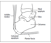
The midfoot is the middle region of the foot, where a cluster of small bones forms an arch on the top of the foot. From this cluster, five long bones (metatarsals) extend to the toes. The bones are held in place by connective tissues (ligaments) that stretch both across and down the foot. However, there is no connective tissue holding the first metatarsal to the second metatarsal. A twisting fall can break or shift (dislocate) these bones out of place.

The Lisfranc joint complex includes the bones and ligaments that connect the midfoot and forefoot. Lisfranc injuries include ligament strains and tears, as well as fractures and dislocations of bone (far right).
The midfoot is critical in stabilizing the arch and in walking (gait). During walking, the midfoot transfers the forces generated by the calf muscles to the front of the foot.
The midfoot joint complex is also called the Lisfranc joint. It is named after French surgeon Jacques Lisfranc de St. Martin, who served in the Napoleonic army in the 1800s.
The Lisfranc joint complex has a specialized bony and ligamentous structure, providing stability to this joint.
Description
The midfoot will be affected if the bones are broken (fractured) or the ligaments are torn (ruptured). Injuries can vary, from a simple injury that affects only a single joint to a complex injury that disrupts multiple different joints and includes multiple fractures.

(Left) This is a subtle injury to the midfoot with widening between the first and second metatarsals (circle), compared with the normal foot on the left. (Center) This x-ray shows a fracture of the second metatarsal (arrow) and a fracture of the cuboid (circle). (Right) This shows a very severe injury of the foot from a high-energy event. It has resulted in a complete dislocation of the entire midfoot (box). Because no bones have been broken, a fusion may be recommended, given the high risk for future arthritis.
Lisfranc injuries tend to damage the cartilage of the midfoot joints. Cartilage covers the ends of bones in the joints, allowing the joints to move smoothly. If severe midfoot injuries are not treated with surgery, then damage to the cartilage and increased stress at the midfoot joints will result in both flatfoot and arthritis, which require complex surgery to correct. Even with successful surgery for the Lisfranc injury, arthritis can still develop in later life.

Cause
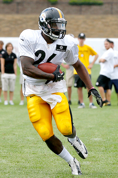
These injuries can happen with a simple twist and fall. This is a low-energy injury. It is commonly seen in football and soccer players. It is often seen when someone stumbles over the top of a foot flexed downwards.
More severe injuries occur from direct trauma, such as a fall from a height. These high-energy injuries can result in multiple fractures and dislocations of the joints.
Symptoms
The most common symptoms of Lisfranc injury include:
  • The top of foot may be swollen and painful.
  • There may be bruising on both the top and bottom of the foot. Bruising on the bottom of the foot is highly suggestive of a Lisfranc injury.
  • Pain that worsens with standing or walking. The pain can be so severe that crutches may be required.
If standard treatment for a sprain (rest, ice, elevation) does not relieve pain and swelling, you should seek care from a Podiatrist at Advanced foot & Ankle Center!.

Doctor Examination

Medical History and Physical Examination

After talking with you about your symptoms and discussing your concerns, your podiatrist will examine your foot and ankle. Although some of the physical tests the podiatrist will perform may be painful, none of them will make the injury worse.

The discoloration on the bottom of the foot is very suggestive of a Lisfranc injury.
Specific things Dr. Raymond A. DiPretoro, Jr. will look for include:
  • Bruising along the bottom of your foot. This suggests a complete tear of the midfoot ligaments or a midfoot fracture.
  • Tenderness to pressure (palpation) along the midfoot.
  • Pain with a stress examination of the midfoot. Dr. DiPretoro may grasp your heel and twist the front of your foot to determine whether there is pain at the midfoot. This should not cause pain in your uninjured foot.
  • Pain with a "piano key" test. Dr. DiPretoro may grasp your toes and move them up and down to determine whether this causes pain. This puts stress across the midfoot and will produce pain if there is an injury.
  • Single limb heel rise. Dr. DiPretoro may ask you to stand on one foot and come up on "tip toes." This places significant stress across the midfoot and is useful if the injury is subtle. This test should not cause pain in your uninjured foot.

Imaging Tests

Other tests that the doctor may order to help confirm your diagnosis include:
X-rays. Broken bones (fractures) and the position of the bones can be seen in an x-ray picture. An x-ray also can show the alignment of the Lisfranc joint. Any change in the normal joint may suggest injury to the ligaments.
If the injury happened after a simple twist and fall (a low-energy injury), the doctor may ask that an x-ray be taken with the patient standing. In this case, the doctor is looking for a ligament injury, especially if the bones are not expected to be broken. Injuries will not be made worse from a standing (weightbearing) x-ray, nor will an injury that might be treated without surgery progress to need surgery if this test is done. The doctor sometimes may take x-rays of your uninjured foot, either for comparison or to determine the stability of the joint.

(Left) In this non-weightbearing x-ray, the Lisfranc injury does not show any abnormal widening (arrow). (Right) The tear of the Lisfranc ligament is more evident in this weightbearing stress x-ray, showing a widening of the joint.
Magnetic resonance imaging (MRI) scan. These studies can create better images of soft tissues like the tendons. This test is not required to diagnose a Lisfranc injury. It may be ordered in cases where the diagnosis may be in doubt.
Computerized tomography scan (CT ) scan. These scans are more detailed than x-rays and can create cross-section images of the foot. This test is not required to diagnose a Lisfranc injury. Because a CT scan will help evaluate the exact extent of the injury and the number of joints that have been injured, a surgeon may order this test to help plan surgery.

Treatment
Treatment for a Lisfranc injury depends on how severe the injury is.

Nonsurgical Treatment

If there are no fractures or dislocations in the joint and the ligaments are not completely torn, nonsurgical treatment may be all that is necessary for healing. A nonsurgical treatment plan includes wearing a non-weightbearing cast for 6 weeks. You must be very strict about not putting weight on your injured foot during this period. This then progresses to weightbearing in a removable cast boot or an orthotic.
Your doctor will want to follow up with you regularly and take additional x-rays to make sure your foot is healing well. In the course of follow up, if there is any evidence that the bones in the injured joint have moved, then surgery will be needed to put the bones back in place.

Surgical Treatment

Surgery is recommended for all injuries with a fracture in the joints of the midfoot or with abnormal positioning (subluxation) of the joints. The goal of surgical treatment is to realign the joints and return the broken (fractured) bone fragments to a normal position.
Internal fixation. In this procedure, the bones are positioned correctly (reduced) and held in place with plates or screws. Because the plates or screws will be placed across joints that normally have some motion, some or all of this hardware may be removed at a later date. This can vary from 3 to 5 months after surgery, and is at the surgeon's discretion.
Occasionally, the hardware may break before it is removed. This is not unusual when screws or plates span bones that have some movement. Metal can fatigue and fail under these conditions, just as a paperclip will fail if bent repeatedly. Most often surgery is successful even if some of the hardware fails.

Various methods of internal fixation can be used to fix Lisfranc injuries. (Left) Multiple screws can be used. (Center) A combination of plates and screws are sometimes required when fractures are present in addition to a torn ligament. (Right) Plates that span the joints are also an excellent method of fixation.
Fusion. If the injury is severe and has damage that cannot be repaired, fusion may be recommended as the initial surgical procedure. A fusion is essentially a "welding" process. The basic idea is to fuse together the damaged bones so that they heal into a single, solid piece.
Lisfranc injuries that may require fusion include joints that cannot be repaired with screws or plates or when the ligaments are severely ruptured. The hardware will not need to be removed because the joints are fused and will not move after they heal.
Rehabilitation. After either surgery (reduction or fusion), a period of nonweightbearing for 6 to 8 weeks is recommended in a cast or cast boot. Weightbearing is started while the patient is in the boot if the x-rays look appropriate after 6 to 8 weeks. The amount of weight a patient can put on their foot, as well as the distance the patient is allowed to walk, is at the surgeon's discretion. Impact activities, such as running and jumping, should be avoided until the hardware has been removed.
Recovery
Some athletes never return to their pre-injury levels of sport after these injuries. Despite excellent surgical reduction and fixation, arthritis may occur from the damage to the cartilage. This may result in chronic pain and may require fusion in the future.

Tuesday, September 17, 2013

Seahawks left tackle, Russell Okung has Turf Toe!


Source: Russell Okung has turf toe

Russell WilsonAP
Seahawks left tackle Russell Okungsustained a turf toe injury in Sunday night’s game vs. San Francisco, a league source told PFT.
Okung departed the game in the first half. He has been replaced by Paul McQuistan.
The 24-year-old Okung was the Seahawks’ No. 1 pick in 2010. He has been the club’s starting left tackle throughout his NFL career.
The Seahawks host the winless Jaguars next Sunday.

Turf Toe: Symptoms, Causes, and Treatments


Turf Toe: Symptoms, Causes, and Treatments

Turf toe is not a term you want to use when talking to a head football coach about his star running back or the ballerina before her diva debut. “Turf toe” is the common term used to describe a sprain of the ligaments around the big toe joint. Although it’s commonly associated with football players who play on artificial turf, it affects athletes in other sports including soccer, basketball, wrestling, gymnastics, and dance.  It’s a condition that’s caused by jamming the big toe or repeatedly pushing off the big toe forcefully as in running and jumping. 
Here is information about turf toe -- what causes it, how to prevent it, and how it's treated -- to help you stay in the game.
football players on field

What Causes Turf Toe?

Turf toe is a sprain to the ligaments around the big toe joint, which works primarily as a hinge to permit up and down motion. Just behind the big toe joint in the ball of your foot are two pea-shaped bones embedded in the tendon that moves your big toe. Called sesamoids, these bones work like a pulley for the tendon and provide leverage when you walk or run. They also absorb the weight that presses on the ball of the foot.
When you are walking or running, you start each subsequent step by raising your heel and letting your body weight come forward onto the ball of your foot. At a certain point you propel yourself forward by "pushing off" of your big toe and allowing your weight to shift to the other foot. If the toe for some reason stays flat on the ground and doesn't lift to push off, you run the risk of suddenly injuring the area around the joint. Or if you are tackled or fall forward and the toe stays flat, the effect is the same as if you were sitting and bending your big toe back by hand beyond its normal limit, causing hyperextension of the toe. That hyperextension, repeated over time or with enough sudden force, can  -- cause a sprain in the ligaments that surround the joint.
Typically with turf toe, the injury is sudden. It is most commonly seen in athletes playing on artificial surfaces, which are harder than grass surfaces and to which cleats are more likely to stick. It can also happen on a grass surface, especially if the shoe being worn doesn't provide adequate support for the foot. Often the injury occurs in athletes wearing flexible soccer-style shoes that let the foot bend too far forward.

What Are the Symptoms of Turf Toe?

The most common symptoms of turf toe include pain, swelling, and limited joint movement at the base of one big toe. The symptoms develop slowly and gradually get worse over time if it’s caused by repetitive injury. If it’s caused by a sudden forceful motion, the injury can be painful immediately and worsen within 24 hours. Sometimes when the injury occurs, a "pop" can be felt. Usually the entire joint is involved, and toe movement is limited.

Monday, September 16, 2013

Sever's Disease (Heel Pain) Exercises and Treatment


What is Sever’s Disease?
Sever's Disease Exercises & Treatment


Sever’s disease is a painful condition of the heel in growing children. The condition occurs as a result of traction on the growth plate at the back of the heel bone where the heel cord (Achilles’ tendon) attaches.

Exercises
Etretching Exercises
The child should stretch his/her heels to provide flexibility. Have your child:


  • Sit in a chair with feet flat on the ground
  • Bend over and grasp his or her toes
  • Pull toes upward until he/she feels a stretch.
  • The child should also stretch out the Achilles’ tendon. Have your child:
  • Stand on the lowest step of the stairs in the house, with only the front part of the feet actually touching the stair.
  • Rise up on tiptoe and then lower himself or herself until the heels are below the level of the step. Use the banister for balance.


Treatment
Treatment

Ice the heel for 15-20 minutes after each practice or game. Insert heel pads in the child’s shoes, and do not let him or her walk around in bare feet. Anti-inflammatory medication (Advil or Aleve) can be used as needed. The pain generally subsides in a few weeks to months, but can last as long as a year. Restriction of activity may be necessary to control symptoms. The exercises above can help the child’s feet stay limber and protect the heels.

Thursday, September 12, 2013

Heel Pain and Sever's Disease in Adolescents!


The calcaneal apophysis is a growth center where the Achilles tendon and the plantar fascia attach to the heel. It first appears in children aged 7 to 8 years. By ages 12 to 14 years the growth center matures and fuses to the heel bone.
Injuries can occur from excessive tension on the Achilles tendon and the plantar fascia, or from direct impact on the heel. Excessive stress on this growth center can cause irritation of the heel, also called Sever’s disease.

Symptoms

Athletes with Sever’s disease are typically aged 9 to 13 years and participate in running or jumping sports such as soccer, football, basketball, baseball, and gymnastics. The typical complaint is heel pain that develops slowly and occurs with activity. The pain is usually described like a bruise. There is rarely swelling or visible bruising. The pain is usually worse with running in cleats or shoes that have limited heel lift, cushion, and arch support. The pain usually goes away with rest and rarely occurs with low-impact sports such as bicycling, skating, or swimming.

Physical exam

A physical exam of the heel will show tenderness over the back of the heel but not in the Achilles tendon or plantar fascia. There may be tightness in the calf muscle, which contributes to tension on the heel. The tendons in the heel get stretched more in patients with flat feet. There is greater impact force on the heels of athletes with a high-arched, rigid foot.

Tests

The doctor may order an x-ray because x-rays can confirm how mature the growth center is and if there are other sources of heel pain, such as a stress fracture or bone cyst. However, x-rays are not necessary to diagnose Sever’s disease, and it is not possible to make the diagnosis based on the x-ray alone.

Other conditions that cause heel pain

Heel pain can also be caused by a stress fracture in the heel, bursitis, tendonitis, bone cysts, and rheumatologic disorders. If the athlete is not active in impact sports or is not between age 9 and 13 years, other conditions should be considered.

Treatment

The following are different treatment options:
  • Rest and modify activity. Limit running and high-impact activity to rest the heel and lessen the pain. Choose one running or jumping sport to play at a time. Substitute low-impact cross-training activities to maintain cardiovascular fitness. This can include biking, swimming, using a stair-climber or elliptical machine, rowing, or inline skating.
  • Reduce inflammation. Ice for at least 20 minutes after activity or when pain increases. Nonsteroidal anti-inflammatory drugs (NSAIDs) may also help.
  • Stretch the calf. Increase calf flexibility by doing calf stretches for 30 to 45 seconds several times per day.

Wednesday, September 11, 2013

We Remember 9/11.....

Just wanted to remember this day by honoring all of those brave people who lost their lives on this horrific day!  We will never forget and will keep you close to our hearts!

We will never forget,

Advanced Foot & Ankle Center


9/11 Memorial Website
September 11, 2001, a day we will never forget
home | 9/11 Websites Other Websites Photos 1 | Photos 2 | Quotes 1 | Quotes 2
 
We recommend this businesses in New York AreaVisit them to help the local economy.
 
Article Hell Directory - An article directory with many great articles.
Education News & Articles - Site offered edcuational articles and latest news.
Hot Traveler Reviews - Offers travel tips, news and articles.

We shall never forget
We shall keep this day,
We shall keep the events and the tears
In our minds, our memory and our hearts
and take them with us as we carry on.
I want to extend appreciation to the people who helped organize events for the September 11, 2001 day. A special thank you to all the people who helped donate and helped contribute to the funds. I want to urge that in your September 11 reflections, you will remember the love, the strength, the hope that carried us through and also brings us together today.
Millions have given time, prayers, blood and money to the relief effort. You may still make a donation to help. Go to the below links.
Some Quotes:

“The US bombing campaign may be hurting coalition friends more than Taliban enemies.” Time, Nov 9.

“Stop this Now.” “Is the war already lost?” New Statesman, Oct 15

“Is bin Laden winning the war?” Spectator Oct 20

[The US forces are] “a gigantic death squad” [and Sept 11 is being] “exploited by capitalists”. Environmentalist George Monbiot in the Guardian

[bin Laden acted from the] “highest motives and in the name of freedom”. Jeanette Winterson, author.

[The Sept 11 attacks were] the greatest work of art in the cosmos … compared to that, we composers are nothing.” Karlheinz Stockhausen, composer. 
``If we learn nothing else from this tragedy, we learn that life is short and there is no time for hate,'' said Sandy Dahl, the wife of Flight 93 pilot Jason Dahl.
Other 9/11 Websites:
September 11, 2001 Memorial
To view more 9/11 Memorial sites please visit our links page.

Some Articles about 9/11:

9/11 HR Hero Gentul Remembered
Born On 9/11
Remembering 9/11, Trying to Prevent a Recurrence
The US militar one year after 9/11
Aviation insurance and 9/11