Thursday, August 29, 2013


What Is Achilles Tendon Inflammation? What Is Achilles Tendinitis?


Achilles tendinitis (tendonitis) or Achilles tendon inflammation occurs when the Achilles tendon becomes inflamed as a result of the Achilles tendon being put under too much strain. The Achilles tendon joins the calf muscles to the heel bone, and is found at the back of a person's lower leg. It is the largest tendon in the body and is able to endure great force, but is still susceptible to injury.

Achiles tendinitis is usually the result of strenuous, high impact exercise, such as running. If ignored, Achilles tendinitis can lead to the tendon tearing or rupturing, and therefore it is important to seek the necessary treatment. Sometimes, treatment can be as simple as getting rest or changing an exercise routine. However, in more severe cases, surgery may be required.

According to Medilexicon's medical dictionary, the Achilles tendon or calcaneal tendon is: 

the thick tendon of insertion of the triceps surae (gastrocnemius and soleus) into the tuberosity of the calcaneus.
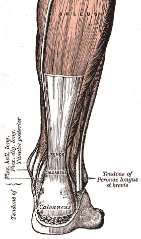

Achilles-tendon
The Achilles tendon (tendo calcaneus) seen from behind 

What are the signs and symptoms of Achilles tendinitis?

A symptom is something the patient feels and reports, while a sign is something other people, such as the doctor, detect. For example, pain is a symptom, while a rash is a sign. The most typical symptom of Achilles tendinitis is a gradual buildup of pain that deteriorates with time. Other possible signs and symptoms of Achilles tendinitis are:
  • the Achilles tendon feels sore a few centimeters above where it meets the heel bone
  • lower leg feels stiff
  • lower leg feels slow and weak
  • slight pain in the back of the leg that appears after running or exercising, and worsens
  • pain in the Achilles tendon that occurs while running or a couple of hours afterwards
  • greater pain experienced when running fast (such as sprinting), for a long time (such as cross country), or even when climbing stairs
  • the Achilles tendon swells or forms a bump
  • the Achilles tendon creaks when touched or moved
Please note that these symptoms, and others similar, can occur in other conditions, so for an accurate diagnosis, a patient would need to visit their doctor.

What are the complications of Achilles tendinitis?

A major complication of Achilles tendinitis is Achilles tendinosis. Achilles tendinosis is a degenerative condition in which the structure of the tendon changes and becomes susceptible to serious damage. This can lead to the tendon tearing and causing great pain. 

When Achilles tendinosis has occurred, and the tendon has torn, surgery would most likely be performed to repair the tendon damage.

What causes Achilles tendinitis?

There are a number of ways a person can develop Achilles tendinitis. Some causes are easier to avoid than others, but being aware of them can aid earlier diagnosis and help prevent serious injury. Causes of Achilles tendinitis include:
  • using incorrect or worn out shoes when running/exercising
  • not warming up properly before exercise
  • increasing intensity of exercise too quickly (e.g. running speed or distance covered)
  • prematurely introducing hill running or stair climbing to exercise routine
  • running on hard/uneven surfaces
  • calf muscle is injured or has little flexibility (this puts a lot of strain on the Achilles tendon)
  • sudden intense physical activity such as sprinting for the finish line
Achilles tendinitis can also be caused by differences in foot, leg or ankle anatomy. For example, some people can have flatness in their foot where there would normally be an arch; this puts more strain on the tendon.

How can Achilles tendinitis be prevented?

Although Achilles tendinitis cannot be completely prevented, the risk of developing it can be lowered. Being aware of the possible causes does help, but the risk can be greatly reduced by taking the following precautions:
  • Getting a variety of exercise - alternating between high-impact exercise (e.g. running) and low-impact exercise (e.g. swimming) can help, as it means there are days when the Achilles tendon is under less tension.
  • Limit certain exercises - doing too much hill running, for example, can put excessive strain on the Achilles tendon.
  • Wearing the correct shoes and replacing them when worn - making sure they support the arch and protect the heel will create less tension in the tendon.
  • Using arch supports inside the shoe - if the shoe is in good condition but doesn't provide the required arch support this is a cheaper (and possibly more effective) alternative to replacing the shoe completely.
  • Stretching - doing this before and after exercising helps to keep the Achilles tendon flexible, which means less chance of tendinitis developing. There is no harm in stretching every day (even on days of rest), as this will only further improve flexibility.
  • Gradually increasing intensity of workout - Achilles tendinitis can occur when the tendon is suddenly put under too much strain, warming up and increasing the level of activity gradually gives your muscles time to loosen up and puts less pressure on the tendon.

How is Achilles tendinitis diagnosed?

When diagnosing Achilles tendinitis, a doctor will ask the patient a few questions about their symptoms and then perform a physical examination. To perform a physical exam on the Achilles tendon, the doctor will lightly touch around the back of the ankle and tendon to locate the source of the pain or inflammation. They will also test the foot and ankle to see if their range of motion and flexibility has been impaired.

The doctor might also order an imaging test to be done on the tendon. This will aid in elimination of other possible causes of pain and swelling, and may help the doctor asses the level of damage (if any) that has been done to the tendon.

Types of imaging tests that could be used for diagnosing Achilles tendinitis are:
  • MRI (Magnetic resonance imaging)
  • X-ray
  • Ultrasound

How can Achilles tendinitis be treated?

The aim when treating Achilles tendinitis is to relieve pain and reduce swelling. The kind of treatment used can vary, based on severity of the condition and whether or not the patient is a professional athlete. After diagnosis, a doctor will decide which method of treatment is required for the patient to undergo, it is likely that they will suggest a combination. Methods used to treat Achilles tendinitis include:
  • Ice packs - applying these to the tendon, when in pain or after exercising, can alleviate the pain and inflammation.
  • Resting - this gives the tissue time to heal. The type of rest needed depends on the severity of the symptoms. In mild cases of Achilles tendinitis, it may mean just reducing the intensity of a workout, in severe cases it might mean complete rest for days or weeks.
  • Elevating the foot - swelling can be reduced if the foot is kept raised above the level of the heart.
  • Exercise and stretching - a doctor might show the patient some stretching exercises that help the Achilles tendon heal, as well as preventing future injury. They may, instead, refer the patient to a physiotherapist or another specialist. The exercises learned will improve flexibility of the area and likely increase calf strength.
  • Pain relievers - non-steroidal anti-inflammatory drugs (NSAIDS), such as ibuprofen can reduce pain and swelling. If you suffer from asthma, kidney disease or liver disease do not take NSAIDs without first checking with your doctor.
  • Steroid injections - these can reduce tendon swelling, but should be performed with caution, as this process has been associated with a greater risk of tendon rupture. A doctor would likely perform the injection while scanning the area with ultrasound to reduce this risk.
  • Compression bandages and orthotic devices - such as ankle supports and shoe inserts can aid recovery, as they take stress off the Achilles tendon.
  • Surgery - as mentioned earlier, surgery is performed to repair any damage to the tendon in cases of Achilles tendinosis. It might also be required if the patient has had recurring Achilles tendinitis and the ongoing inflammation has caused the tendon to rupture. A doctor may also resort to surgery when several of the above treatment methods have proved unsuccessful.

Monday, August 26, 2013

What is Neuropathy? Neuropathy Causes and Treatments


Neuropathy is a collection of disorders that occurs when nerves of the peripheral nervous system (the part of the nervous system outside of the brain and spinal cord) are damaged. The condition is generally referred to as peripheral neuropathy, and it is most commonly due to damage to nerve axons. Neuropathy usually causes pain and numbness in the hands and feet. It can result from traumatic injuries, infections, metabolic disorders, and exposure to toxins. One of the most common causes of neuropathy is diabetes.


Neuropathy can affect nerves that control muscle movement (motor nerves) and those that detect sensations such as coldness or pain (sensory nerves). In some cases - autonomic neuropathy - it can affect internal organs, such as the heart, blood vessels, bladder, or intestines.
Pain from peripheral neuropathy is often described as a tingling or burning sensation. There is no specific length of time that the pain exists, but symptoms often improve with time - especially if the neuropathy has an underlying condition that can be cured. The condition is often associated with poor nutrition, a number of diseases, and pressure or trauma, but many cases have no known reason (called idiopathic neuropathy).
In the United States, about 20 million people suffer from neuropathy. Over half of diabetes patients also suffer from the condition.

How is neuropathy classified?

Peripheral neuropathy can be broadly classified into the following categories:
  • Mononeuropathy - involvement of a single nerve. Examples include carpal tunnel syndrome, ulnar nerve palsy, radial nerve palsy, and peroneal nerve palsy.
  • Multiple mononeuropathy - two or more nerves individually affected.
  • Polyneuropathy - generalized involvement of peripheral nerves. Examples include diabetic neuropathy and Guillain-Barre syndrome. 
Neurophathies may also be categorized based on a functional classification (motor, sensory, autonomic, or mixed) or the type of onset (acute - hours or days, subacute - weeks or months, or chronic - months or years).
The most common form of neuropathy is (symmetrical) peripheral polyneuropathy, which mainly affects the feet and legs on both sides of the body.

What causes neuropathy?

About 30% of neuropathy cases are considered idiopathic, which means they are of unknown cause. Another 30% of neuropathies are due to diabetes. In fact, about 50% of people with diabetes develop some type of neuropathy. The remaining cases of neuropathy, called acquired neuropathies, have several possible causes, including:
  • Trauma or pressure on nerves, often from a cast or crutch or repetitive motion such as typing on a keyboard
  • Nutritional problems and vitamin deficiencies, often from a lack of B vitamins
  • Alcoholism, often through poor dietary habits and vitamin deficiencies
  • Autoimmune diseases, such as lupus, rheumatoid arthritis, and Guillain-Barre syndrome
  • Tumors, which often press up against nerves
  • Other diseases and infections, such as kidney disease, liver disease, Lyme disease, HIV/AIDS, or an underactive thyroid (hypothyroidism)
  • Inherited disorders (hereditary neuropathies), such as Charcot-Marie-Tooth disease and amyloid polyneuropathy
  • Poison exposure, from toxins such as heavy metals, and certain medications and cancer treatments

Who gets neuropathy?

Risk factors for peripheral neuropathy include several conditions and behaviors. People with diabetes who poorly control their blood sugar levels are very likely to suffer from some neuropathy. Autoimmune diseases such as lupus and rheumatoid arthritis also increase one's chance of developing a neuropathy. People who have received organ transplants, AIDS patients, and others who have had some type of immune system suppression have a higher risk of neuropathy. In addition, those who abuse alcohol or have vitamin deficiencies (especially B vitamins) are at an increased risk. Neuropathy is also more likely to occur in people with kidney, liver or thyroid disorders.

Thursday, August 22, 2013

Overlapping Toes


Description


Deformities of the toes are common in the pediatric population. Generally they are congenital in nature with both or one of the parents having the same or similar condition. Many of these deformities are present at birth and can become worse with time. Rarely do children outgrow these deformities although rare instances of spontaneous resolution of some deformities have been reported.
Malformation of the toes in infancy and early childhood are rarely symptomatic. The complaints of parents are more cosmetic in nature. However as the child matures these deformities progress from a flexible deformity to a rigid deformity and become progressively symptomatic. Many of these deformities are unresponsive to conservative treatment. Common digital deformities are underlapping toesoverlapping toesflexed or contracted toes and mallet toes. Quite often a prolonged course of digital splitting and exercises may be recommended but generally with minimal gain. As the deformity becomes more rigid surgery will most likely be required if correction of the deformity is the goal.

Underlapping Toes

Description

Underlapping toes are commonly seen in the adult and pediatric population. The toes most often involved are the fourth and fifth toes. A special form of underlapping toes is called clinodactyly or congenital curly toes. Clinodactyly is fairly common and follows a familial pattern. One or more toes may be involved with toes three, four, and five of both feet being most commonly affected.
The exact cause of the deformity is unclear. A possible etiology is an imbalance in muscle strength of the small muscles of the foot. This is aggravated by a subtle abnormality in the orientation on the joints in the foot just below the ankle joint called the subtalar joint. This results in an abnormal pull of the ligaments in the toes causing them to curl. With weight bearing the deformity is increased and a folding or curling of the toes results in the formation of callous on the outside margin of the end of the toe. Tight fitting shoes can aggravate the condition.

Treatment

The age of the patient, degree of the deformity and symptoms determine treatment. If symptoms are minimal, a wait and see approach is often the best bet. When treatment is indicated the degree of deformity determines the level of correction. When the deformity is flexible in nature a simple release of the tendon in the bottom of the toe will allow for straightening of the toe. If the deformity is rigid in nature then removal of a small portion of the bone in the toe may be necessary. Both of these procedures are common in the adult patient for the correction of hammertoe deformity. If skin contracture is present a derotational skin plasy may be required.

Overlapping Toes - Overlapping Fifth Toe

Description

This deformity is characterized by one toe lying on top of an adjacent toe. The most common toe involved is the fifth toe. When one of the central toes is involved the second toe is most commonly affected. The etiology of the condition is not well understood. It is though that it may be caused by the position of the fetus in the womb during development. The condition my run in families so there may be a hereditary component to the deformity.

Treatment

Effective conservative treatment depends upon how early the diagnosis is made. In infancy, passive stretching and adhesive tapping is most commonly used. This may require 6 to 12 weeks to accomplish and reoccurrence is not uncommon. Rarely will the deformity correct itself. As the individual matures the deformity becomes fixed. When surgical correction is warranted a skin plasty is required to release the contracture of the skin associated with the deformity. Additionally a tendon release and a release of the soft tissues about the joint at the base of the fifth toe may be required. In severe cases the toe may require the placement of a pin to hold the toe in a straighten position. The pin, which exits the tip of the toe, may be left in place for up to three weeks. During this period of time the patient must curtail their activities significantly and wear either a post-operative type shoe or a removable cast. Excessive movement at the surgical site can result in a less than desirable result. The pin can be easily removed in the doctor's office with minimal discomfort. Following removal of the pin splinting of the toe may be required for an additional two to three weeks.

Hammertoes and Mallet Toes

Description

Another common digital deformity is contracture of the toes in the formation of hammertoes and mallet toes. Hammertoes are described in depth in another article. Mallet toes are a result of contracture of the last joint in the toe. In the pediatric population it is often flexible and not painful. Over time the deformity becomes rigid and a callous may form on the skin overlying the joint at the end of the toe. Additionally the toenail may become thickened and deformed form the repetitive jamming of the toe while walking. The deformity usually involves one or two toes, with the second toe most commonly affected. Mallet toes have several etiologies. Longer toes that are forced against a short toe box in the shoe will, over time, develop a contracture of the last joint in the toe causing a mallet toe.

Treatment

Conservative treatment consists of padding and strapping the toes into a corrected position. This treatment may alleviate the symptoms but will not correct the deformity. Diabetic patients often develop ulcerations on the ends of their toes secondary to mallet toe deformity and the pressure that results from the toe jamming into the shoe. When standing, the toe will demonstrate a contracture, with the tip of the toe facing downward into the floor. If the deformity is flexible a simple release of the tendon in the bottom of the toe will allow straightening of the toe. Following the procedure however the patient must avoid shoes that cause jamming of the toe or the deformity can reoccur. When the deformity is rigid surgical correction requires the removal of a small section of bone in the last joint of the toe. On occasion fusion of the last two bones in the toe may be necessary. This requires removing the cartilage from the last joint in the toe and pinning the bones together. When the bone heals it forms a single bone and the toe remains in a straightened position. Healing time is dependent upon the procedure selected. If a tendon release is performed the patient my return to a roomy shoe within a week. If the toe is straightened by removing a section of the bone in the toe it make ten days to three weeks for a patient to return to normal shoes. If a fusion is performed to straighten the toe, the patient may not return to normal shoes for 6 to 8 weeks. Time off from work will depend upon the type of shoe gear that must be worn and the level of activity necessary to perform the job. A minimum of three to four days off from work is generally recommended and longer if the job responsibilities can not be modified to accommodate the normal healing time for the surgery.

Do You Have Arch Pain?


Definition 

The term arch pain (often referred to as arch strain) refers to an inflammation and/or burning sensation at the arch of the foot.

Cause

There are many different factors that can cause arch pain. A structural imbalance or an injury to the foot can often be the direct cause. However, most frequently the cause is a common condition called plantar fasciitis. The plantar fascia is a broad band of fibrous tissue located along the bottom surface of the foot that runs from the heel to the forefoot. Excessive stretching of the plantar fascia, usually due to over-pronation (flat feet), causes plantar fasciitis. The inflammation caused by the plantar fascia being stretched away from the heel often leads to pain in the heel and arch areas. The pain is often extreme in the morning when an individual first gets out of bed or after a prolonged period of rest. If this condition is left untreated and strain on the longitudinal arch continues, a bony protrusion may develop, known as a heel spur. It is important to treat the condition promptly before it worsens.

Treatment and Prevention

This is a common foot condition that can be easily treated. If you suffer from arch pain avoid high-heeled shoes whenever possible. Try to choose footwear with a reasonable heel, soft leather uppers, shock absorbing soles and removable foot insoles. When the arch pain is pronation related (flat feet), an orthotic designed with a medial heel post and proper arch support is recommended for treating the pain. This type of orthotic will control over-pronation, support the arch and provide the necessary relief.

If the problem persists, consult your Podiatrist at Advanced Foot & Ankle Center!


Do You Have Calluses?


Definition

The formation of calluses is caused by an accumulation of dead skin cells that harden and thicken over an area of the foot. This callus formation isthe body's defense mechanism to protect the foot against excessive pressure and friction. Calluses are normally found on the ball-of-the-foot, the heel, and/or the inside of the big toe. Some calluses have a deep seated core known as a nucleation. This particular type of callus can be especially painful to pressure. This condition is often referred to as Intractable Plantar Keratosis.

Cause

Calluses develop becuase of excessive pressure at a specific area of the foot. Some common causes of callus formation are high-heeled dress shoes, shoes that are too small, obesity, abnormalities in the gait cycle (walking motion), flat feet, high arched feet, bony prominences, and the loss of the fat pad on the bottom of the foot.

Treatment and Prevention

Many people try to alleviate the pain caused by calluses by cutting or trimming them with a razor blade or knife. This is not the way to properly treat calluses. This is very dangerous and can worsen the condition resulting in unnecessary injuries. Diabetics especially should never try this type of treatment. To relieve the excessive pressure that leads to callus formation, weight should be redistributed equally with the use of an orthotic. An effective orthotic transfers pressure away from the "hot spots" or high pressured areas to allow the callus to heal. The orthotic should be made with materials that absorb shock and shear (friction) forces. Women should also steer away from wearing high-heeled shoes. As always, surgery should be the very last resort.

If the problem persists, consult your Podiatrist at Advanced Foot and Ankle Center!


Wednesday, August 21, 2013

Interesting foot Facts!


Foot Facts


  • A quarter of all the body’s bones are in the feet (There are 52 bones in a pair of feet).
  • The average child will take its first steps around 13-17 months - but between 10 and 18 months falls within the “normal” range. 
  • During the first year of a child’s life their feet grow rapidly, reaching almost half their adult size. By 12, a child’s foot is about 90 per cent of its adult length. 
  • When walking, each time your heel lifts off the ground it forces the toes to carry one half of your body weight. 
  • It’s rare that two feet are exactly the same; one of them is often larger than the other. 
  • In a pair of feet, there are 250,000 sweat glands. 
  • The first foot coverings were probably animal skins, which Stone Age peoples in northern Europe and Asia tied around their ankles in cold weather. 
  • Cigarette smoking is the biggest cause of Peripheral Vascular Disease (disease of the arteries of the feet and legs) which often leads to pain on walking, ulceration, infection and in the most severe cases - gangrene and possible amputation. 
  • Foot disorders in the elderly are extremely common and are the cause of much pain and disability, and consequent loss of mobility and independence. 
  • A human foot & ankle is a strong, mechanical structure that contain 26 bones, 33 joints, and more than 100 muscles, tendons & ligaments. 
  • During an average day of walking, the total forces on your feet can total hundreds of tons, equivalent to an average of a fully loaded cement truck. 
  • Walking is the best exercise for your feet. It contributes to your general health by improving circulation and weight control. 
  • Standing in one spot is far more tiring than walking because the demands are being made on the same few muscles for a longer length of time. 
  • Foot ailments can become your first sign of more serious medical problems. Your feet mirror your general health, so conditions like arthritis, diabetes, nerve and circulatory disorders can show their initial symptoms in your feet. 
  • 75% of Americans will experience foot problems at one time or another in their lives. 
  • Butterflies taste with their feet, gannets incubate eggs under their webbed feet and elephants use their feet to hear – they pick up vibrations of the earth through their soles. 
  • Sweat glands in the feet produce approximately half a pint of perspiration daily. 
  • The average person will walk around 115,000 miles in a life time, that's more than four times around the earth! 
  • 3 out of 4 Americans experience serious foot problems in their lifetime. 
  • The foot contains 26 bones, 33 joints, 107 ligaments and 19 muscles. 
  • 1/4 of all the bones in the human body are down in your feet. When these bones are out of alignment, so is the rest of the body. 
  • Only a small percentage of the population is born with foot problems. 
  • It's neglect and a lack of awareness of proper care - including ill fitting shoes - that bring on problems. 
  • Women have about four times as many foot problems as men. High heels are partly to blame. 
  • Walking is the best exercise for your feet. It also contributes to your general health by improving circulation, contributing to weight control, and promoting all-around well being. 
  • Your feet mirror your general health. Conditions such as arthritis, diabetes, nerve and circulatory disorders can show their initial symptoms in the feet - so foot ailments can be your first sign of more serious medical problems. 
  • Arthritis is the number one cause of disability in America. It limits everyday dressing, climbing stairs, getting in and out of bed or walking - for about 7 million Americans. 
  • About 60-70% of people with diabetes have mild to severe forms of diabetic nerve damage, which in severe forms can lead to lower limb amputations. Approximately 56,000 people a year lose their foot or leg to diabetes. 
  • There are 250,000 sweat glands in a pair of feet. Sweat glands in the feet excrete as much as a half-pint of moisture a day. 
  • Walking barefoot can cause plantar warts. The virus enters through a cut. 
  • The two feet may be different sizes. Buy shoes for the larger one. 
  • About 5% of Americans have toenail problems in a given year. 
  • The average person takes 8,000 to 10,000 steps a day, which adds up to about 115,000 miles over a lifetime. That's enough to go around the circumference of the earth four times. 
  • There are currently more websites on the Internet having to do with foot fetishes than with foot health. 
  • The record for the world’s largest feet belongs to Matthew McGrory who wears US size 28 1/2 shoes. The average men’s size is 10 1/2. 
  • Madeline Albrecht holds the world record for most feet sniffed at 5,600. 
  • The foot accounts for 25% of the bones in the human body. 
  • The average woman walks three miles more per day than the average male. 
  • There are roughly 250,000 sweat glands on a pair of feet. 
  • Sweat glands in the feet produce as much as half a pint of moisture each day. 
  • The pressure on the feet when running can be as much as four times the runners body weight. 
  • The first shoes were invented 5,000,000 years ago during the Ice Age and were made from animal skins. 
  • The afternoon is the optimum time to shop for shoes because the feet tend to be more swollen then. 
  • 9 out of 10 women wear shoes that are too small for them. 
  • Women experience foot problems four times more than men. 
  • Fingernails and toenails grow faster during hot weather, pregnancy and teenage years. 
  • The ancient Romans were the first to construct distinct left and right shoes. Before that, shoes could be worn on either foot. 
  • The average foot gets two sizes longer when a person stands up. 
  • Shoe sizes were devised in England by King Edward II who declared in 1324 that the diameter of one barely corn- a third of an inch- would represent one full shoe size. That’s still true today. 
  • The feet can contract an array of nasty diseases from communal showers: Planter Wart, Athletes foot, Ring worm! 

Diabetes-Foot Ulcers

Diabetes-Foot Ulcers


Bookmark & Share
If you have diabetes, you may have an increased risk for developing foot sores, or ulcers. Foot ulcers are the most common reason for hospital stays for people with diabetes. It may take weeks or even several months for your foot ulcers to heal. Diabetic ulcers are often painless.
Whether or not you have a foot ulcer, you will need to learn more about taking care of your feet.

Debridement

Debridement is the process to remove dead skin and tissue. Your Podiatrist will need to do this to be able to see your foot ulcer. There are many ways to do this.
One way is to use a scalpel and special scissors.
  • The skin surrounding the wound is cleaned and disinfected.
  • The wound is probed with a metal instrument to see how deep it is and to see if there is any foreign material or object in the ulcer.
  • Dr. Raymond A. DiPretoro, Jr., cuts away the dead tissue, then washes out the ulcer.
  • Your sore may seem bigger and deeper after the doctor or nurse debrides it. The ulcer should be red or pink in color and look like fresh meat.
Other ways to remove dead or infected tissue are to:
  • Put your foot in a whirlpool bath.
  • Use a syringe and catheter (tube) to wash away dead tissue.
  • Apply wet to dry dressings to the area to pull off dead tissue.
  • Put special chemicals, called enzymes, on your ulcer. These dissolve dead tissue from the wound.

Taking Pressure off of Your Foot Ulcer

Foot ulcers are partly caused by too much pressure on one part of your foot. Be sure to wear shoes that do not put a lot of pressure on your foot. Try not to have pressure over the ulcer too.
Your Podiatrist at Advanced Foot & Ankle Center may ask you to wear special shoes, or a brace or a special cast. You may need to use a wheelchair or crutches for awhile. These devices will take the pressure off of the ulcer area. This will help speed up the healing process.
The type of shoes you wear when you have diabetes is important:
  • Wear shoes made out of canvas, leather, or suede. Do not wear shoes made out of plastic, or other material that does not breathe.
  • Wear shoes you can adjust easily. They should have laces, Velcro, or buckles.
  • Wear shoes that fit properly and have plenty of room in them. You may need a special shoe made to fit your foot.
  • Do not wear shoes with pointed or open toes, such as high heels, flip-flops, or sandals.

Wound Care and Dressings

You will need to do these things to care for your wound:
  • Keep your blood sugar levels under tight control. This will help you heal faster.
  • Keep the ulcer clean and bandaged.
  • Cleanse the wound daily, using a wound dressing or bandage.
  • Try to take fewer steps around your house.
  • Do not walk barefoot unless your Podiatrist tells you it is OK.
Your Podiatrist may use different kinds of dressings to treat your ulcer.
Wet-to-dry dressings are often used first. This process involves applying a wet dressing to your wound. As the dressing dries, it absorbs wound material. When the dressing is removed, some of the tissue comes off with it.
  • Your Podiatrist will tell you how often you need to change the dressing.
  • You may be able to change your own dressing, or family members may be able to help.
  • A visiting nurse may also help you.
Other types of dressings are:
  • Dressing that contain calcium alginates or growth factors
  • Skin substitutes
Keep your dressing and the skin around it dry. Try not to get healthy tissue around your wound too wet from your dressings. This can soften the healthy tissue and cause more foot problems.

When to Call your Podiatrist

Call your Podiatrist at Advanced Foot & Ankle Center if you have any of these signs and symptoms of infection:
  • Redness, increased warmth, or swelling around the wound
  • Extra drainage
  • Pus
  • Odor
  • Fever or chills
  • Increased pain
  • Increased firmness around the wound
Also call your Podiatrist if your foot ulcer is very white, blue, or black.

Friday, August 16, 2013

Diabetes, Foot Care and Foot Ulcers!


Diabetes, Foot Care and Foot Ulcers


Some people with diabetes develop foot ulcers. A foot ulcer is prone to infection, which may become severe. This leaflet aims to explain why foot ulcers sometimes develop, what you can do to help prevent them, and typical treatments if one does occur.
A skin ulcer is where an area of skin has broken down and you can see the underlying tissue. Most skin ulcers occur on the lower legs or feet. The skin normally heals quickly if it is cut. However, in some people with diabetes the skin on the feet does not heal so well and is prone to developing an ulcer. This can be even after a mild injury such as stepping on a small stone in your bare feet.
Foot ulcers are more common if you have diabetes because one or both of the following complications develop in some people with diabetes:


Reduced sensation of the skin on your feet

Your nerves may not work as well as normal because even a slightly high blood sugar level can, over time, damage some of your nerves. This is a complication of diabetes called peripheral neuropathy of diabetes.

The nerves that take messages of sensation and pain from the feet are commonly affected. If you lose sensation in parts of your feet, you may not know if you damage your feet. For example, if you tread on something sharp or develop a blister due to a tight shoe. This means that you are also more prone to problems such as minor cuts, bruises or blisters. Also, if you cannot feel pain so well from the foot, you do not protect these small wounds by not walking on them. Therefore, they can quickly become worse and develop into ulcers.

Narrowing of arteries (blood vessels) going to the feet

If you have diabetes you have an increased risk of developing narrowing of the arteries (peripheral vascular disease). This is caused by fatty deposits called atheroma that build up on the inside lining of arteries (sometimes called furring of the arteries). This can reduce the blood flow to various parts of the body.

The arteries in the legs are quite commonly affected. This can cause a reduced blood supply (poor circulation) to the feet. Skin with a poor blood supply does not heal as well as normal and is more likely to be damaged. Therefore, if you get a minor cut or injury, it may take longer to heal and be prone to becoming worse and developing into an ulcer. In particular, if you also have reduced sensation and cannot feel the wound.


  • If you have reduced sensation to your feet (see above). The risk of this occurring increases the longer you have diabetes and the older you are. Also, if your diabetes is poorly controlled. This is one of the reasons why it is very important to keep your blood sugar level as near normal as possible.
  • If you have narrowed arteries (see above). The risk of this occurring increases the longer you have diabetes, the older you become and also if you are male. Also, if you have any other risk factors for developing furring of the arteries. For example, if you smoke, do little physical activity, have a high cholesterol level, high blood pressure or are overweight.
  • If you have had a foot ulcer in the past.
  • If you have other complications of diabetes, such as kidney or eye problems.
  • If your feet are more prone to minor cuts, grazes, corns or calluses which can occur:
    • If you have foot problems such as bunions which put pressure on points on the feet.
    • If your shoes do not fit properly, which can put pressure on your feet.
    • If you have leg problems which affect the way that you walk, or prevent you from bending to care for your feet.
Although foot ulcers can be serious, they usually respond well to treatment. However, foot ulcers can get worse and can take a long time to heal if you have diabetes, particularly if your circulation is not so good. In addition, having diabetes means you are more likely to have infections and an infection in the ulcer can occur. Occasionally, more serious problems can develop, such as gangrene.


Have your feet regularly examined

Most people with diabetes are reviewed at least once a year by Dr. Raymond A. DiPretoro, Jr. and other health professionals. Part of this review is to examine your feet to look for problems such as reduced sensation or poor circulation. If any problems are detected then more frequent feet examinations will usually be recommended.

Treatment of diabetes and other health risk factors

As a rule, the better the control of your diabetes, the less likely you are to develop complications such as foot ulcers. Also, where appropriate, treatment of high blood pressure, high cholesterol level and reducing any other risk factors will reduce your risk of diabetic complications. In particular, if you smoke, you are strongly advised to stop smoking.


Foot care

Research has shown that people with diabetes who take good care of their feet and protect their feet from injury, are much less likely to develop foot ulcers.

Good foot care includes:
  • Looking carefully at your feet each day, including between the toes. If you cannot do this yourself, you should get someone else to do it for you:
    • Looking is particularly important if you have reduced sensation in your feet, as you may not notice anything wrong at first until you look.
    • If you see anything new (such as a cut, bruise, blister, redness or bleeding) and don't know what to do, see your doctor or podiatrist (chiropodist).
    • Do not try to deal with corns, calluses, verrucas or other foot problems by yourself. They should be treated by a Podiatrist at Advanced Foot and Ankle Center!  In particular, do not use chemicals or acid plasters to remove corns, etc.
    • Use a moisturising oil or cream for dry skin to prevent cracking. However, you should not apply it between the toes as this can cause the skin to become too moist which can lead to an infection developing.
    • Look out for athlete's foot (a common minor skin infection). It causes flaky skin and cracks between the toes, which can be sore and can become infected. If you get athlete's foot, it should be treated with an antifungal cream.
  • Cut your nails by following the shape of the end of your toe. But, do not cut down the sides of the nails, or cut them too short, or use anything sharp to clean down the sides of the nails. These things may cause damage or lead the nail to develop an ingrown nail. If you cannot see properly do not try to cut your nails, as you may cut your skin. You should ask someone else to do it.
  • Wash your feet regularly and dry them carefully, especially between the toes.
  • Do not walk barefoot, even at home. You might tread on something and damage your skin.
  • Always wear socks with shoes or other footwear. However, don't wear socks that are too tight around the ankle, as they may affect your circulation.
  • Shoes, trainers and other footwear should:
    • Fit well to take into account any awkward shapes or deformities (such as bunions).
    • Have broad fronts with plenty of room for the toes.
    • Have low heels to avoid pressure on the toes.
    • Have good laces, buckles or Velcro® fastening to prevent movement and rubbing of feet within the shoes.
  • When you buy shoes, wear the type of socks that you usually wear. Avoid slip-on shoes, shoes with pointed toes, sandals and flip-flops. Break in new shoes gradually.
  • Always feel inside footwear before you put footwear on (to check for stones, rough edges, etc).
  • If your feet are an abnormal shape, or if you have bunions or other foot problems, you may need specially fitted shoes to stop your feet rubbing.
  • Tips to avoid foot burns include: checking the bath temperature with your hand before stepping in; do not use hot water bottles, electric blankets or foot spas; do not sit too close to fires.
You should tell your Podiatrist, Dr. Raymond A. DiPretoro, Jr. right away if you suspect an ulcer has formed. Treatment aims to dress and protect the ulcer, to prevent or treat any infection and also to help your skin to heal.
  • The ulcer is usually covered with a protective dressing.
  • Dr. DiPretoro will normally examine, clean and re-dress the ulcer regularly.
  • Dr. DiPretoro may need to remove any hard skin that prevents the ulcer from healing. Also, depending on the site and size of the ulcer, they may protect it from further injury by using padding to take the pressure off the area.
  • You may also be advised to wear special shoes or have a cast made for your foot to keep the pressure off the ulcer.
  • Antibiotics will be advised if the ulcer, or nearby tissue, becomes infected.
  • Sometimes a small operation is needed to drain pus and clear dead tissue if infection becomes more severe.
  • In some cases, the arteries in the legs are very narrow and greatly reduce the blood flow to the feet. In these cases an operation to bypass, or widen, the arteries may be advised.
Many foot ulcers will heal with the above measures. However, they can take a long time to heal.

In some cases, the ulcer becomes worse, badly infected and does not heal. Sometimes infection spreads to nearby bones or joints, which can be difficult to clear, even with a long course of antibiotics. Occasionally, the tissue in parts of the foot cannot survive and the only solution then is to amputate the affected part.


If you have a diabetic foot problem, you will be able to get most of the treatment you need from your GP or other health professionals working in the community. However, there are some problems which may require you to go into hospital for treatment. The National Institute for Health and Clinical Excellence (NICE) has released some guidance as to what you can expect if this should happen:
  • You should expect to be treated by a team which deals especially with people who have diabetic foot problems. This team usually includes a diabetic specialist, a surgeon who deals with diabetic foot problems, a podiatrist, a diabetes nurse specialist and a tissue viability nurse (who assesses whether the skin and underlying tissues of your feet have been affected by circulation changes due to diabetes).
  • You should be given information about your foot condition.
  • You will have your diabetes assessed and checks will be made to make sure you have not developed complications such as kidney disease (or if you have already developed complications, to make sure they are not getting any worse).
  • You will have an examination of both feet to check for ulcers, cuts and abrasions to the feet, signs of poor circulation, areas of numbness and the development of Charcot's arthropathy. This is a condition in which the sensation of pain is reduced because of the diabetes, resulting in damage to bones, such as tiny fractures.
  • You will have a general examination to make sure you do not have a fever or any other signs of a severe generalised infection.
  • If you have an ulcer, this will be checked for infection and baseline measurements of the size and depth will be taken.
  • You may be asked to have an X-ray or scan of your foot to make sure the bones have not been affected by your condition.

Diabetic Wound Care


Diabetic Wound Care

A diabetic foot ulcer is an open sore or wound that occurs in approximately 15 percent of patients with diabetes and is commonly located on the bottom of the foot. Of those who develop a foot ulcer, 6 percent will be hospitalized due to infection or other ulcer-related complication.
Diabetes is the leading cause of non-traumatic lower extremity amputations in the United States, and approximately 14-24 percent of patients with diabetes who develop a foot ulcer will require an amputation. Foot ulceration precedes 85 percent of diabetes-related amputations. Research has shown, however, that development of a foot ulcer is preventable.  
Causes
Anyone who has diabetes can develop a foot ulcer. Native Americans, African Americans, Hispanics, and older men are more likely to develop ulcers. People who use insulin are at higher risk of developing a foot ulcer, as are patients with diabetes-related kidney, eye, and heart disease. Being overweight and using alcohol and tobacco also play a role in the development of foot ulcers.  
Ulcers form due to a combination of factors, such as lack of feeling in the foot, poor circulation, foot deformities, irritation (such as friction or pressure), and trauma, as well as duration of diabetes. Patients who have diabetes for many years can develop neuropathy, a reduced or complete lack of ability to feel pain in the feet due to nerve damage caused by elevated blood glucose levels over time. The nerve damage often can occur without pain, and one may not even be aware of the problem. Your podiatrist can test feet for neuropathy with a simple, painless tool called a monofilament.
Vascular disease can complicate a foot ulcer, reducing the body's ability to heal and increasing the risk for an infection. Elevations in blood glucose can reduce the body's ability to fight off a potential infection and also slow healing. 
Symptoms
Because many people who develop foot ulcers have lost the ability to feel pain, pain is not a common symptom. Many times, the first thing you may notice is some drainage on your socks. Redness and swelling may also be associated with the ulceration and, if it has progressed significantly, odor may be present.
When to Visit a Podiatrist
Once an ulcer is noticed, call Advanced Foot and Ankle Center immediately. Foot ulcers in patients with diabetes should be treated to reduce the risk of infection and amputation, improve function and quality of life, and reduce health-care costs.
Diagnosis and Treatment
The primary goal in the treatment of foot ulcers is to obtain healing as soon as possible. The faster the healing, the less chance for an infection.
There are several key factors in the appropriate treatment of a diabetic foot ulcer:
  • Prevention of infection
  • Taking the pressure off the area, called “off-loading”
  • Removing dead skin and tissue, called “debridement”
  • Applying medication or dressings to the ulcer
  • Managing blood glucose and other health problems
Not all ulcers are infected; however, if your podiatrist diagnoses an infection, a treatment program of antibiotics, wound care, and possibly hospitalization will be necessary.
To keep an ulcer from becoming infected, it is important to:
  • keep blood glucose levels under tight control;
  • keep the ulcer clean and bandaged;
  • cleanse the wound daily, using a wound dressing or bandage; and
  • avoid walking barefoot.
For optimum healing, ulcers, especially those on the bottom of the foot, must be “off-loaded.”  You may be asked to wear special footgear, or a brace, specialized castings, or use a wheelchair or crutches.  These devices will reduce the pressure and irritation to the area with the ulcer and help to speed the healing process.
The science of wound care has advanced significantly over the past ten years. The old thought of “let the air get at it” is now known to be harmful to healing. We know that wounds and ulcers heal faster, with a lower risk of infection, if they are kept covered and moist. The use of full-strength betadine, hydrogen peroxide, whirlpools, and soaking are not recommended, as these practices could lead to further complications.
Appropriate wound management includes the use of dressings and topically-applied medications. Products range from normal saline to growth factors, ulcer dressings, and skin substitutes that have been shown to be highly effective in healing foot ulcers.
For a wound to heal, there must be adequate circulation to the ulcerated area.  Dr. Raymond A. DiPretoro, Jr. can determine circulation levels with noninvasive tests. 
Tightly controlling blood glucose is of the utmost importance during the treatment of a diabetic foot ulcer. Working closely with a medical doctor or endocrinologist to control blood glucose will enhance healing and reduce the risk of complications.
Surgical Options: A majority of non-infected foot ulcers are treated without surgery; however, if this treatment method fails, surgical management may be appropriate. Examples of surgical care to remove pressure on the affected area include shaving or excision of bone(s) and the correction of various deformities, such as hammertoes, bunions, or bony “bumps.”
Healing time depends on a variety of factors, such as wound size and location, pressure on the wound from walking or standing, swelling, circulation, blood glucose levels, wound care, and what is being applied to the wound. Healing may occur within weeks or require several months.
Prevention
The best way to treat a diabetic foot ulcer is to prevent its development in the first place. Recommended guidelines include seeing a podiatrist on a regular basis. Dr. Raymond A. DiPretoro, Jr. can determine if you are at high risk for developing a foot ulcer and implement strategies for prevention.
You are at high risk if you have or do the following:
  • Neuropathy
  • Poor circulation
  • A foot deformity (e.g., bunion, hammer toe)
  • Wear inappropriate shoes
  • Uncontrolled blood sugar
  • History of a previous foot ulceration
Reducing additional risk factors, such as smoking, drinking alcohol, high cholesterol, and elevated blood glucose, are important in prevention and treatment of a diabetic foot ulcer. Wearing the appropriate shoes and socks will go a long way in reducing risks.  Advanced Foot and Ankle Center can provide guidance in selecting the proper shoes.
Learning how to check your feet is crucial so that you can find a potential problem as early as possible. Inspect your feet every day—especially the sole and between the toes—for cuts, bruises, cracks, blisters, redness, ulcers, and any sign of abnormality. Each time you visit a health-care provider, remove your shoes and socks so your feet can be examined. Any problems that are discovered should be reported to Dr. DiPretoro as soon as possible; no matter how simple they may seem to you. 
The key to successful wound healing is regular podiatric medical care to ensure the following “gold standard” of care:
  • Lowering blood sugar
  • Appropriate debridement of wounds
  • Treating any infection
  • Reducing friction and pressure
  • Restoring adequate blood flow